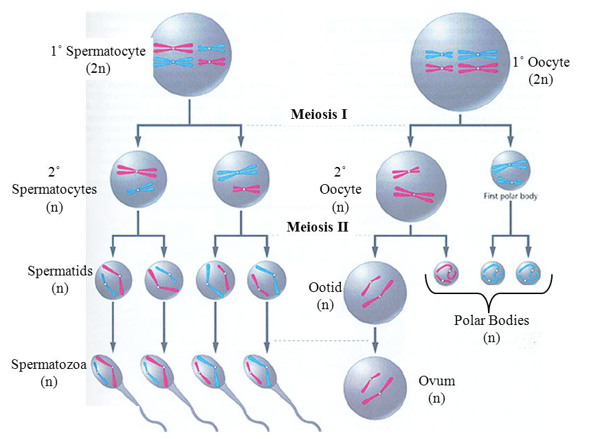
Meiosis in Males (spermatogenesis)
Meiosis 1
During Meiosis 1, homologous chromosomes are separated.
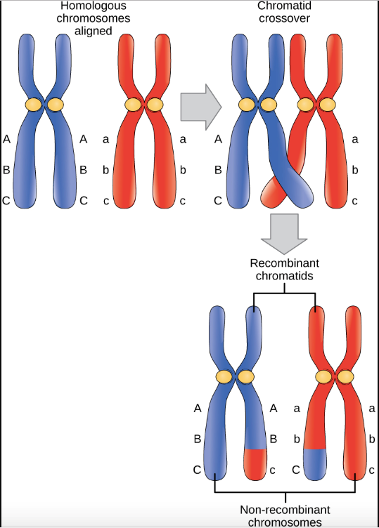
Prophase 1: The homologous chromosomes come together and cross over to form a new combination of DNA on each chromosome.
Metaphase 1: The chromosomes attach to spindle fibres at the kinetochore. The chromosomes line up at the middle of the cells. This arrangement of paired chromosomes and their formation at the middle is random which gives rise to the genetic variation. The amount of variation is over 8 million different combinations.
Anaphase 1:
The chromosomes that are paired then begin to separate as they are pulled apart by the spindle fibres.
Telophase 1:
The chromosomes reach opposite ends of the cell. A membrane starts to form between the two side of the cell.
Cytokinesis:
The cell has divided. There is one copy of each chromosome in the 2 new cells.
Meiosis 2 in males: The goal is to separate each chromosome into two chromatids
Prophase 2: Chromosomes begin to condense. The nuclear membrane begins to dissolve, and the spindle fibres begin to form.
Metaphase 2: Spindle fibres attach to the chromosomes. The chromosomes are lined up at the centre of the cell.
Anaphase 2: sister chromatids are pulled apart by the spindle fibres. Sister chromatids are separated and move to opposite side of cell.
Telophase 2: Chromosomes have move to the opposite pole of the cell. Nuclear membrane forms around the chromosomes.
Cytokinesis: Cells are completely separated, and cell division is done. These cells will develop further into a mature sperm cell.
Meiosis in females (oogenesis): During Meiosis 1, homologous chromosomes are separated.
Prophase 1: The homologous chromosomes come together and cross over to form a new combination of DNA on each chromosome.
Metaphase 1: The chromosomes attach to spindle fibres at the kinetochore. The chromosomes line up at the middle of the cells. This arrangement of paired chromosomes and their formation at the middle is random which gives rise to the genetic variation. The amount of variation is over 8 million different combinations.
Anaphase 1: The chromosomes that are paired then begin to separate as they are pulled apart by the spindle fibres.
Telophase 1: The chromosomes reach opposite ends of the cell. A membrane starts to form between the two side of the cell. In females, one cell between the two sister cells becomes the polar body and one becomes the egg cell precursor. The polar cell is small, and the egg cell is large. This is to allow the egg cell to survive.
Cytokinesis: The cell has divided. A polar body cell and an egg cell precursor have one copy of each chromosome.
Meiosis 2 in females: The goal is to separate each chromosome into two chromatids
Prophase 2: Chromosomes begin to condense. The nuclear membrane begins to dissolve, and the spindle fibres begin to form.
Metaphase 2: Spindle fibres attach to the chromosomes. The chromosomes are lined up at the centre of the cell.
Anaphase 2: sister chromatids are pulled apart by the spindle fibres. Sister chromatids are separated and move to opposite side of cell.
Telophase 2: Chromosomes have move to the opposite pole of the cell. Nuclear membrane forms around the chromosomes.
Cytokinesis: Cells are completely separated, and cell division is done. 3 polar bodies are formed and one egg cell.
References:
"The Cell Cycle, Mitosis and Meiosis." University of Leicester. August 17, 2017. Accessed June 22, 2018. https://www2.le.ac.uk/projects/vgec/highereducation/topics/cellcycle-mitosis-meiosis.
Bryant, Lindsay. "Gametogenesis the Formation of Gametes in Animals Gametogenesis Although Gametogenesis in Males and Females Is Generally the Same There Are Some General." SlidePlayer. Accessed June 22, 2018. http://slideplayer.com/slide/9675145/.
Genetic Variation and Diversity in Meiosis
There are 2 sources of variation that occurs during meiosis:
Independent random assortment of chromosomes is a factor in genetic variation during the metaphase stages of meiosis. In this picture, we can see that in possibility 1, there are 2 combinations that can occur. The maternal (blue) and paternal (red) genes are separated into separate cells since they are randomly arranged to be on opposite sides of the original cell. In the second possibility, the maternal and paternal genes are on both sides of the cell (maternal/paternal on top and paternal/maternal on bottom). This results in combinations 3 and 4. This is a simplification of the random assortment process showing only 4 pairs of chromosomes. In humans, there are 23 pairs of chromosomes so the possibilities of different combinations during the Metaphase I is over 8 million (2^23). This amount of diversity does not even consider the second factor of genetic variation called ‘crossover.’

The second form of variation is crossover. This occurs when the 2 pairs of chromosomes crossover during metaphase and line up at the centre of the cell. Once the chromatid strands from the chromosomes have crossed over, then they are pulled apart which causes the crossed over pieces to be exchanged as seen in the picture below. This results in a new formation of genetic sequence resulting in vast diversity.
References:
"Biology for Majors I." Lumen. Accessed June 28, 2018. https://courses.lumenlearning.com/wm-biology1/chapter/reading-genetic-variation-in-meiosis/.
"Brent Cornell." Intestinal Villi | BioNinja. Accessed June 28, 2018. http://ib.bioninja.com.au/higher-level/topic-10-genetics-and-evolu/101-meiosis/random-assortment.html.
https://cnx.org/resources/63f8c2e1463100032093c794742bcd9a/Figure_11_01_02.jpg